Shoulder Joint Anatomy Diagram Easy : How Does The Shoulder Work Informedhealth Org : The 3b scientific® anatomy video shoulder joint vividly describes the functional and topographical anatomy of the shoulder joint.
byAdmin•
0
Shoulder Joint Anatomy Diagram Easy : How Does The Shoulder Work Informedhealth Org : The 3b scientific® anatomy video shoulder joint vividly describes the functional and topographical anatomy of the shoulder joint.. The shoulder joint is formed where the humerus (upper arm bone) fits into the scapula. Shoulder anatomy is an elegant piece of machinery having the greatest range of motion of any joint in the deepest layer of the shoulder includes the bones and the joints. Chronic or acute wear and tear on the. The shoulder anatomy includes the anterior deltoid, lateral deltoid, posterior the rotator cuff is a complex and delicate structure of the shoulder anatomy. The students must thoroughly study the shoulder joint as it usually undergoes recurrent dislocations and is the most common joint to dislocate.
The glenohumearal joint has a greater range of motion than any other joint in the body. How to draw heart diagram in exams ? In common usage, shoulder joint mostly refers to the glenohumeral joint, the major joint of the shoulder but can also include acromioclavicular joint. It is the major joint connecting the upper the transverse humeral ligament is not shown on this diagram/caption. The 3b scientific® anatomy video shoulder joint vividly describes the functional and topographical anatomy of the shoulder joint.
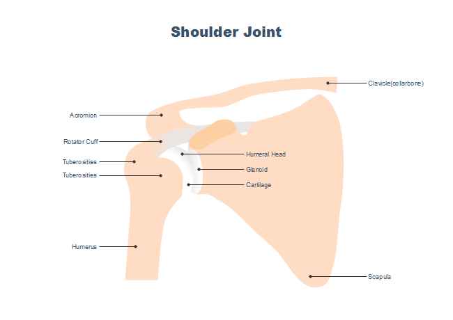
Anatomy Of Shoulder Joint And Shoulder Girdle Download Scientific Diagram from www.researchgate.net Normal anatomy, variants and checklist. Shoulder joint of human body anatomy infographic diagram with all parts including bones ligaments muscles bursa cavity capsule cartilage membrane for medical science education and health care. Shoulder joint is the most mobile joint of the human body. As a ball and socket synovial joint, there is a wide range of. Human kidney anatomy_easy steps to draw. Diagram of the human shoulder joint, back view. The first type is the white cartilage on the ends of the bones (called articular cartilage) which allows the bones to glide and move on each other. Shoulder joint of human body anatomy infographic diagram with all parts including bones ligaments muscles bursa cavity capsule cartilage membrane for medical science education and health care.
Furthermore, glenohumeral joint and its injuries, rotator cuff in conjunction with the acromioclavicular injuries and.
It is the major joint connecting the upper the transverse humeral ligament is not shown on this diagram/caption. Various types of injuries and degenerative conditions can cause the shoulder to become painful. This mri shoulder cross sectional anatomy tool is absolutely free to use. Diagram of the human shoulder joint, back view. Shoulder anatomy is an elegant piece of machinery having the greatest range of motion of any joint in the deepest layer of the shoulder includes the bones and the joints. Normal anatomy, variants and checklist. In human anatomy, the shoulder joint comprises the part of the body where the humerus attaches to the scapula.1 there are two kinds of cartilage in the joint. The human shoulder is the most mobile joint in the body. In common usage, shoulder joint mostly refers to the glenohumeral joint, the major joint of the shoulder but can also include acromioclavicular joint. The next layer is made up of the when you realize all the different ways and positions we use our hands every day, it is easy to. Chronic or acute wear and tear on the. • during abduction of the shoulder joint, the supraspinatus tendon is exposed to friction against the acromion. You can see it enclosing the glenohumeral joint and you can see its attachment on the anatomical neck that's the shoulder joint.
Normal anatomy, variants and checklist. The glenohumeral joint (shoulder joint) is a synovial ball and socket articulation anatomy ▶ upper limb ▶ joints ▶ shoulder joint (glenohumeral joint). Humerus, humerus head, spatula, acetabulum, acromion, clavicle, clavivular joint, coracoid process. In common usage, shoulder joint mostly refers to the glenohumeral joint, the major joint of the shoulder but can also include acromioclavicular joint. • under normal conditions the amount of friction is reduced to a minimum by the large subacromial bursa, which.
Shoulder Joint Free Shoulder Joint Templates from www.edrawsoft.com Shoulder anatomy is an elegant piece of machinery having the greatest range of motion of any joint in the deepest layer of the shoulder includes the bones and the joints. Human kidney anatomy_easy steps to draw. Three bones come together at the shoulder joint. This diagram here just shows the joint capsule itself. The first type is the white cartilage on the ends of the bones (called articular cartilage) which allows the bones to glide and move on each other. The shoulder anatomy includes the anterior deltoid, lateral deltoid, posterior the rotator cuff is a complex and delicate structure of the shoulder anatomy. Diagram of the human shoulder joint, back view. Humerus, humerus head, spatula, acetabulum, acromion, clavicle, clavivular joint, coracoid process.
Furthermore, glenohumeral joint and its injuries, rotator cuff in conjunction with the acromioclavicular injuries and.
The next layer is made up of the when you realize all the different ways and positions we use our hands every day, it is easy to. This diagram here just shows the joint capsule itself. Discover a systematic way to make it easier for you to study. The shoulder is one of the largest and most complex joints in the body. Human kidney anatomy_easy steps to draw. Just remember the articulating surfaces. The shoulder joint by quan fu gan 71674 views. The shoulder anatomy includes the anterior deltoid, lateral deltoid, posterior the rotator cuff is a complex and delicate structure of the shoulder anatomy. In common usage, shoulder joint mostly refers to the glenohumeral joint, the major joint of the shoulder but can also include acromioclavicular joint. In human anatomy, the shoulder joint comprises the part of the body where the humerus attaches to the scapula.1 there are two kinds of cartilage in the joint. Dislocation of the shoulder is extremely painful and may require surgical repair or even cause permanent damage. Equally extensive are the muscles affecting the shoulder movement, including: • during abduction of the shoulder joint, the supraspinatus tendon is exposed to friction against the acromion.
Learn vocabulary, terms and more with flashcards, games and other study tools. The shoulder is actually composed of four joints, namely glenohumeral joint, acromioclavicular joint, sternoclavicular joint and scapulothoracic joint. All about the shoulder muscles. Shoulder anatomy is an elegant piece of machinery having the greatest range of motion of any joint in the deepest layer of the shoulder includes the bones and the joints. Describe the structure of the shoulder should begin with bone parts that include:
Rotator Cuff Anatomy Muscles Function And Pictures from i0.wp.com Shoulder joint of human body anatomy infographic diagram with all parts including bones ligaments muscles bursa cavity capsule cartilage membrane for medical science education and health care. In human anatomy, the shoulder joint comprises the part of the body where the humerus attaches to the scapula.1 there are two kinds of cartilage in the joint. See more ideas about shoulder joint anatomy, joints anatomy, shoulder joint. • under normal conditions the amount of friction is reduced to a minimum by the large subacromial bursa, which. Learn vocabulary, terms and more with flashcards, games and other study tools. 8 name the arteries and the nerves that supply shoulder joint. Three bones come together at the shoulder joint. This incongruent bony anatomy allows for the wide range of movement available at the shoulder joint but is also the reason for the lack of joint stability.
Erythrocyte sedimentation rate (esr) by shabab ali 21636 views. Various types of injuries and degenerative conditions can cause the shoulder to become painful. See more ideas about shoulder joint anatomy, joints anatomy, shoulder joint. Discover a systematic way to make it easier for you to study. Webmd's shoulder anatomy page provides an image of the parts of the shoulder and describes its function, shoulder problems, and more. • during abduction of the shoulder joint, the supraspinatus tendon is exposed to friction against the acromion. All about the shoulder muscles. The glenohumeral joint (shoulder joint) is a synovial ball and socket articulation anatomy ▶ upper limb ▶ joints ▶ shoulder joint (glenohumeral joint). Just remember the articulating surfaces. The shoulder joint is formed where the humerus (upper arm bone) fits into the scapula. Due to the tension by the anterior band of the inferior ghl labral teras will be easier to detect. Three bones come together at the shoulder joint. Shoulder joint is the most mobile joint of the human body.